
Breathing is a function we perform about twenty thousand times a day without thinking about it. Most of the time, our body takes in only the air it needs to maintain baseline functions at rest. When we climb a steep staircase carrying heavy luggage, however, or when we attempt to run, we immediately realize that our respiratory system has reserves that remain idle during sedentary life. This range between the minimum and maximum amount of air we can move is called functional lung capacity.
How, though, can we calculate how much air our chest actually holds without opening up the body? The lungs are not like rigid containers such as a glass bottle; instead, they resemble two elastic sponges that fill and empty within a moving cage of ribs and muscles. Measuring this capacity in practice is not just a metric for athletes, but a fundamental tool showing how freely the airways move and how effectively the muscular pump of the diaphragm works.
What are vital capacity and tidal volume?
To understand the measurements, we must first divide lung air into separate compartments. When you sit quietly in a chair reading, you inhale and exhale a small amount of air. This amount is called tidal volume. You can picture it as a 500-milliliter (ml) glass of water that you continually empty and refill in a bucket. The body does not exhaust its space with every breath; it moves only the oxygen it needs at that moment to avoid tiring the respiratory muscles.
What happens, though, if you take the deepest breath possible and then empty all the air until nothing is left? This maximum volume you can voluntarily move is called vital capacity (VC). Vital capacity includes the resting tidal volume, plus the extra air you can forcibly draw in, plus the air you can force out after a normal exhalation. It represents the full working range of your pulmonary system during intense physical exertion.
There is, however, a volume that cannot be measured with a simple exhalation. This is called residual volume (RV). It is the amount of air that necessarily remains in the alveoli even after the most forceful exhalation. Without this air, the delicate lung tissues would stick together like wet plastic bags and would require immense muscular pressure to reopen on the next breath. The sum of vital capacity and residual volume gives us total lung capacity.
| Respiratory volume | What it represents | Typical value in a healthy adult |
|---|---|---|
| Tidal volume (TV) | The air of a quiet, automatic breath. | 400 to 600 ml |
| Inspiratory reserve volume (IRV) | The additional air that fits after a normal inhalation. | 2000 to 3000 ml |
| Expiratory reserve volume (ERV) | The additional air that is expelled after a normal exhalation. | 1000 to 1500 ml |
| Residual volume (RV) | The air that never exits, preventing lung collapse. | 1100 to 1400 ml |
| Vital capacity (VC) | The total air moved from maximum inhalation to maximum exhalation. | 3500 to 5000 ml |
How a medical spirometer works
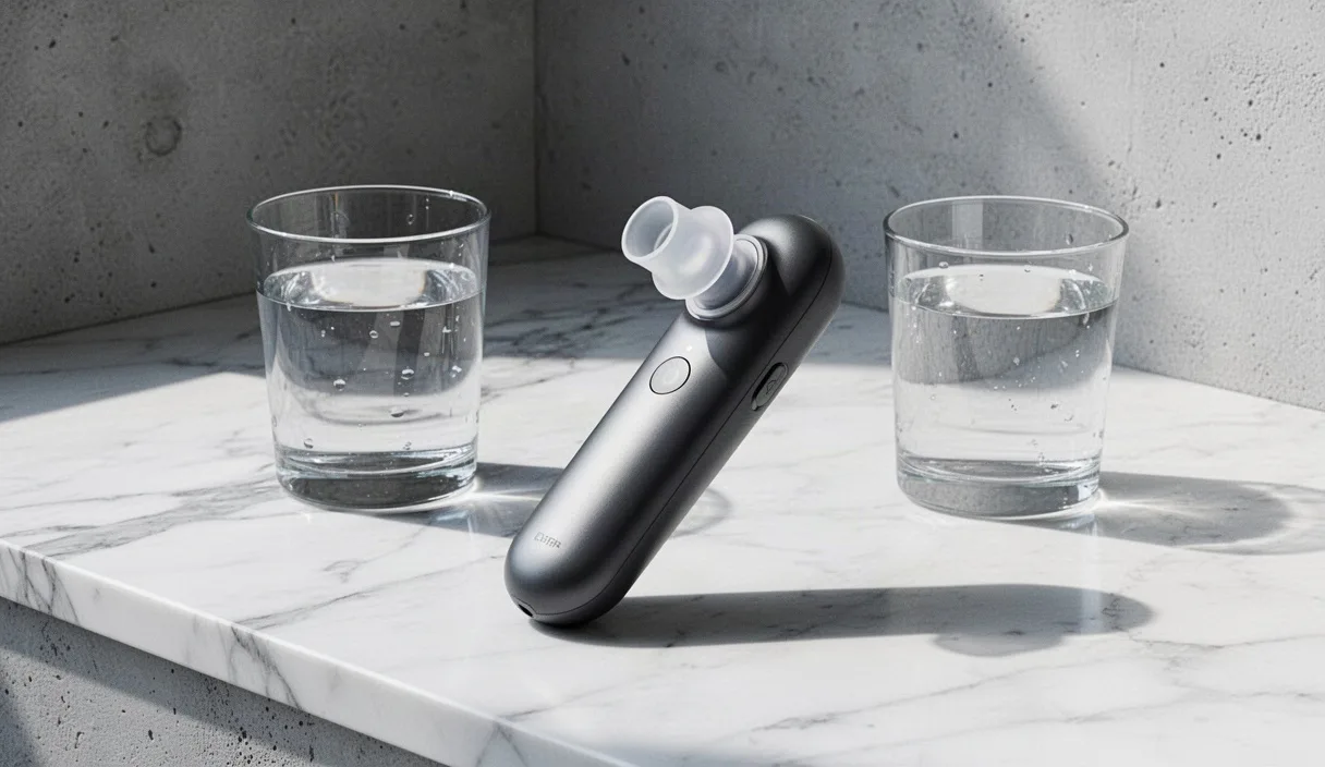
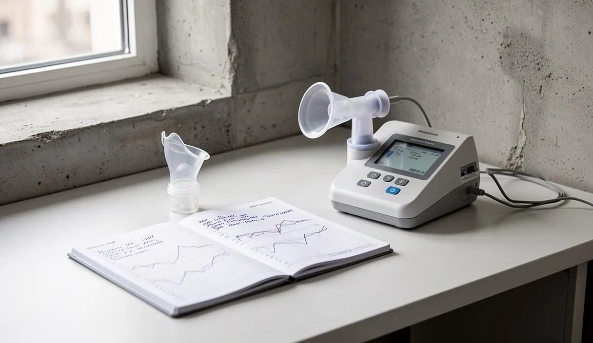
In clinical practice, the primary instrument that converts breathing into measurable graphs is the spirometer. The device consists of a disposable mouthpiece, a flow sensor, and an electronic recording system. During the test, the patient wears a soft nose clip so that all air passes exclusively through the mouthpiece. The sensor usually contains a tiny turbine or a pressure difference membrane. As the air moves, the turbine spins, and its rotation speed translates directly into flow rate and volume per fraction of a second.
The procedure requires intense effort and proper guidance from the healthcare professional. The patient sits with a straight back, takes a deep, full inhalation expanding their chest to the maximum limit, seals their lips around the mouthpiece, and exhales with as much force and speed as possible, continuing to blow for at least six seconds, even when feeling completely empty. This test is usually repeated at least three times to ensure results are consistent and reliable.
From this test, the physician derives two critical values:
- FVC (Forced Vital Capacity): The total volume of air in liters that the patient was able to expel during this forceful exhalation.
- FEV1 (Forced Expiratory Volume in 1 second): The amount of air expelled exclusively during the first second of the effort.
Why does the first second matter? Think of two 2-liter bottles filled with water. One has a wide opening and empties almost immediately when turned upside down. The other has a narrow straw. Both contain the same amount, but the second one takes much longer to empty. In the human body, if total air volume (FVC) is normal but the amount exhaled in the first second is low, this indicates to specialists that the airways are narrowed or partially blocked, as seen in asthma or chronic obstructive pulmonary disease.
Which values are considered expected based on height and age
There is no single number that applies to everyone. A person who is two meters tall has a larger rib cage and consequently larger lungs than someone who is one meter sixty centimeters tall. For a physician to determine whether a result is satisfactory, the spirometer does not compare the patient to the general population average, but to international reference tables (such as Global Lung Function Initiative data) corresponding to the same sex, same age, and same exact height.
How does age affect the lungs? Just as skin gradually loses its elasticity over the years, the same happens with lung tissue and rib joints. After the age of 25 to 30, when the development of the respiratory system is complete, vital capacity naturally declines by about 25 to 35 milliliters per year. This natural aging process means that an FVC value of 3.8 liters might be excellent for a 65-year-old man, but considered low for a 22-year-old athlete of the exact same height.
| Individual Profile | Height | Indicative Expected FVC | Indicative Expected FEV1 |
|---|---|---|---|
| Male, 25 years old | 182 cm | 5.10 to 5.50 liters | 4.20 to 4.60 liters |
| Male, 65 years old | 182 cm | 3.90 to 4.30 liters | 3.00 to 3.40 liters |
| Female, 25 years old | 165 cm | 3.40 to 3.80 liters | 2.90 to 3.30 liters |
| Female, 65 years old | 165 cm | 2.60 to 3.00 liters | 2.00 to 2.40 liters |
Typically, a measurement is considered within normal ranges when it is above 80% of the predicted value for the individual's specific demographic characteristics, while the FEV1 to FVC ratio in healthy young adults generally ranges between 75% and 85%. If someone experiences symptoms of shortness of breath, only a pulmonologist can interpret these deviations in conjunction with medical history.
Can an adult increase lung size?
The straightforward anatomical answer is no. After adolescence ends and physical growth is finalized, the number of alveoli in the lungs stabilizes around 300 to 480 million. The rib cage no longer grows taller, and the lungs do not gain new tissue. An adult who starts training will not increase the physical volume of their organs the way a bicep grows in the gym.
Why, then, do endurance athletes and swimmers seem to breathe with such ease? What changes significantly with exercise is not the size of the lungs, but the efficiency of breathing mechanics and the endurance of respiratory muscles. The diaphragm, the primary dome-shaped muscle separating the chest from the abdomen, becomes stronger and more agile. The intercostal muscles learn to expand the ribs using less energy, allowing the person exercising to approach their theoretical maximum vital capacity without fatiguing.
Furthermore, regular aerobic activity improves blood circulation around the alveoli and increases the heart's pumping capacity. This enables the body to absorb more oxygen from the exact same volume of air. Thus, while the anatomical size of the lungs remains constant, their functional utilization shifts from a shallow, incomplete movement into a deep, economical, and coordinated function.
Simple breath observation tests without equipment
Although no home method can replace the precision of a medical spirometer, there are simple observational tests you can perform at home. These tests do not serve as a medical diagnosis, but they offer insight into the control, mobility, and awareness of your respiratory system.
Measuring resting respiratory rate
The rate at which you breathe when not exerting yourself shows how efficiently your body manages its oxygen requirements. To measure it properly:
- Sit comfortably in an armchair and remain still for at least five minutes to allow your heart rate to drop.
- Place a timer in front of you. Do not try to control your breathing; let it flow naturally.
- Count how many times your chest or abdomen rises over the course of one full minute (one inhalation and one exhalation equal one respiratory cycle).
A typical resting rate for a healthy adult ranges between 12 and 18 breaths per minute. Values consistently above 20 breaths in a state of complete relaxation usually indicate shallow, inefficient breathing or muscular tension.
Checking chest expansion with a tape measure
This test shows how freely your ribs move and how much room they allow for the lungs to expand.
- Stand upright in front of a mirror and wrap a soft sewing tape measure around your chest, right at the base of the sternum (below the armpits).
- Exhale all air slowly and steadily. At the end of the exhalation, record the measurement in centimeters without overtightening the tape.
- Take a maximum deep breath, expanding your ribs outward to the sides, and record the new measurement.
In healthy adults, the difference between full exhalation and full inhalation usually ranges between 4 and 7 centimeters. A difference of less than 3 centimeters often indicates stiffness of the rib cage or a habit of shallow breathing using only the upper chest.
Timed controlled exhalation
This test evaluates your ability to regulate airflow using your respiratory muscles. Take a full, comfortable breath through your nose and exhale through your mouth with your lips pursed into a very small opening, as if gently blowing on a candle flame without extinguishing it. Count the seconds until your smooth airflow ends. A duration of 20 to 35 seconds indicates good diaphragmatic control and adequate reserve volume.
Common measurement and assessment mistakes
In both informal checks and clinical tests, several common errors lead to misleading conclusions about lung health:
- Poor lip seal on the spirometer: If you leave even a tiny gap at the corners of your mouth during a forceful exhalation, air escapes into the room. This falsely reduces the recorded FVC value, creating the impression that your lungs are smaller.
- Ending the exhalation too early: Many people stop blowing after three seconds because they feel pressure in their chest. However, smaller airways continue emptying until the sixth or seventh second. Stopping prematurely cuts off the measurement of true lung volume.
- Confusing breath-holding with capacity: How long you can hold your breath underwater depends mainly on your brain's tolerance to carbon dioxide, not on the size of your lungs. A person with lower vital capacity may hold their breath longer than someone with large lungs simply because they tolerate the urge to breathe better.
- Incorrect posture: Leaning forward while exhaling compresses abdominal organs against the diaphragm. An accurate measurement always requires an upright, lengthened spine so the diaphragm has full downward freedom of motion.
Practical daily steps
If you want to maintain your current lung capacity at the best possible level and support your respiratory health, there are specific actions you can add to your routine:
- Schedule a medical exam if symptoms are present: If you notice unexplained shortness of breath during simple daily tasks, chronic coughing, or wheezing, visit a pulmonologist for a formal spirometry test. Home checks cannot rule out underlying conditions.
- Incorporate 30 minutes of moderate aerobic exercise into your day: Brisk walking, swimming, or cycling encourages the respiratory system to work close to its tidal volume limits, preserving thoracic flexibility.
- Practice diaphragmatic breathing for five minutes: Lie on your back with your knees bent and one hand resting on your stomach. Inhale through your nose, making sure only the hand on your abdomen rises, not your chest. This trains your primary breathing muscle to use its full range of motion.
- Improve your workstation ergonomics: Prolonged sitting with rounded shoulders compresses the chest cavity and restricts natural lung expansion. Position your monitor at eye level and take short rib-stretching breaks every hour.